

Репозитарій ПВНЗ "ХММУ"
ПРИВАТНИЙ ВИЩИЙ НАВЧАЛЬНИЙ ЗАКЛАД «ХАРКІВСЬКИЙ МІЖНАРОДНИЙ МЕДИЧНИЙ УНІВЕРСИТЕТ»
Електронний репозитарій (архів) накопичує, зберігає та надає вільний доступ у режимі онлайн до електронних версій наукових, навчальних та науково-методичних праць спільноти університету.
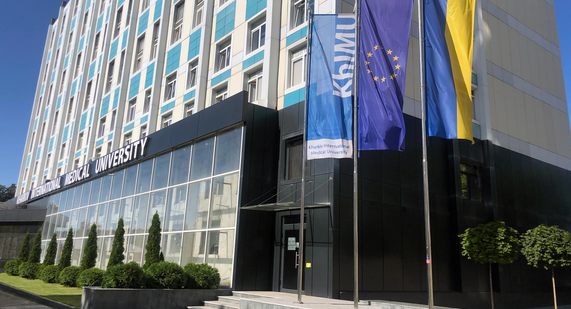
KhIMURep – (Kharkiv International Medical University Repository) – відкритий електронний архів (репозитарій) ПВНЗ «Харківський міжнародний медичний університет».
Фонди
Виберіть фонд, щоб переглянути його зібрання.
- Офіційні наукові видання Харківського міжнародного медичного університету: матеріали конференцій, форуми, збірники наукових праць університету. Публікації співробітників у матеріалах конференцій інших установ шукайте в розділі "Наукові праці дослідників університету".
- Наукові публікації, статті, монографії та дослідження професорсько-викладацького складу, науковців та здобувачів ХММУ
Нові надходження
Item type:Документ, Захворювання слизової оболонки порожнини рота у дитячому віці(Харків : Харківський національний університет імені В. Н. Каразіна, 2026) Ніконов, Андрій Юрійович; Фоменко, Юлія Володимирівна; Іскоростенська, Ольга Володимирівна; Щебликіна, Наталія Альбертівна; Колесова, Тамара Олексіївна; Назарян, Розана Степанівна; Ткаченко, Марина Вікторівна; Nikonov, Andriy Yuriyovych; Fomenko, Yuliia Volodymyrivna; Iskorostenska, Olha Volodymyrivna; Shcheblykina, Nataliia Albertivna; Kolesova, Tamara Oleksiyivna; Nazaryan, Rozana Stepanivna; Tkachenko, MarynaНавчально-методичний посібник містить розділи терапевтичної стоматології дитячого віку, що вивчають особливості будови і функції слизової оболонки порожнини рота у дітей. У посібнику стисло надано характеристику основних захворювань слизової оболонки порожнини рота у дитячому віці та висвітлено методи діагностики і лікування. Слизова оболонка порожнини рота виконує велику кількість клінічно значущих функцій. Вона діє як захисний фізичний та імунний бар’єр від зовнішніх подразників, підтримує рівень вологості тканини за рахунок функціонування слинних залоз, а також сприймає і реагує на температурні, больові та сенсорні подразнення. Численні захворювання можуть вражати слизову оболонку порожнини рота та погіршувати її здатність виконувати ці завдання. Поширені поверхневі ураження слизової оболонки порожнини рота, спричинені вірусами або грибами, проявляються рецидивуючим герпесом, кандидозом або рецидивуючими стоматитами. Наприклад, пошкодження епітелію порожнини рота може спричинити порушення клітинної диференціації та виникнення запальних або пухлинних процесів. Крім того, руйнування ясеневого бар’єру, спричинене дисбактеріозом мікробіоти порожнини рота, призводить до захворювань пародонту. Патологічні стани слизової оболонки порожнини рота у дітей можуть бути проявом при соматичних та інфекційних захворюваннях дитячого віку. Варто зазначити, що патологія слизової оболонки порожнини рота може бути симптомом багатьох системних захворювань. Тому знання гістологічних особливостей слизової оболонки порожнини рота є обов’язковим для розпізнавання та точної діагностики патологічних доброякісних та злоякісних новоутворень у дитячому віці. The guide contains sections on pediatric dentistry, studying the features of the oral structure and function in children. The guide summarizes the underlying oral disease in childhood and highlights the diagnostic and treatment methods. The oral mucosa performs different clinically relevant functions. It physically and immunologicaly protects the oral cavity from exogenous irritants, maintains the tissue moisture level due to the functioning of the salivary glands, and responds to temperature, pain, and sensory stimulation. Numerous diseases can affect the oral mucosa and impair its ability to perform these functions. Common superficial oral lesions caused by viruses or fungi are manifested by recurrent herpes, candidiasis, or recurrent stomatitis. For example, oral epithelium damage can lead to impairment of cellular differentiation and inflammation or neoplastic processes. In addition, breaking the gingival barrier caused by oral microbiota dysbiosis leads to periodontal disease. Oral mucosal disease in children can be manifested by pediatric somatic and infectious disease. It is worth noting that the oral mucosal disease can be a symptom of many systemic diseases. Therefore, histological characterization of the oral mucosa is mandatory for the detection and accurate diagnosis of benign and malignant neoplasms in childhood.Item type:Документ, Аспекти використання інформаційних технологій у підготовці майбутніх медичних фахівців(2026) Коваленко, Наталія Іллівна; Ткаченко, Марина ВікторівнаАктуальність. Трансформація сучасної вищої медичної освіти вимагає активного впровадження цифрових інструментів для формування цифрової компетентності здобувачів та забезпечення високої якості практичної підготовки. Мета дослідження. Вивчення функціональних можливостей сучасних електронних ресурсів і обґрунтування їхньої ефективності під час дистанційного навчання здобувачів вищої медичної освіти. Методи та зміст. Проаналізовано досвід застосування симуляційних методів, 3D-графіки, віртуальної (VR), доповненої (AR) та імерсивної віртуальної реальності (IVR), систем із тактильним/гаптичним зворотним зв’язком (VR-Haptic: Simodont Dental Trainer, Unidental, HapTEL), а також інструментів штучного інтелекту у вищій медичній школі. Результати. Застосування зазначених технологій дозволяє моделювати складні біологічні та медичні процеси, вивчати рідкісні патології за базами МРТ-знімків, а також безпечно відпрацьовувати мануальні навички й техніку хірургічних чи стоматологічних втручань без використання трупного матеріалу та етичних ризиків для пацієнтів. Висновки. Інтеграція VR/AR-платформ та штучного інтелекту збагачує інтерактивні методи навчання, сприяє розвитку просторового мислення, засвоєнню теоретичних знань і практичних навичок, підвищуючи загальну якість освітнього процесу в медичних закладах.Item type:Документ, Рентгеноморфометричне прогнозування виникнення нових компресійних переломів тіл хребців після вертебропластики(2026) Попов, А. І.; Молодюк, М. В.; Куценко, В. О.; Златнік, Р. В.; Нессонова, М. М.Актуальність. Нові компресійні переломи хребців (НКПХ) після пункційної вертебропластики (ПВП) є серйозною проблемою вертебрології. Методи. У ретроспективне когортне дослідження включено 26 пацієнтів (24 жінки, 2 чоловіки; середній вік $(69,04 \pm 2,04)$ року) з остеопоротичними компресійними переломами хребців, яким виконувалася ПВП. Вивчено морфометричні параметри 99 хребців на цифрових рентгенограмах (RadiAnt DICOM Viewer, точність $0,1\text{ мм} / 0,1^\circ$): висоти $h_a$ (передня), $h_m$ (середня), $h_p$ (задня), відносна компресія $H_a, H_m, H_p$ (%), індекс клиноподібності (ІК), локальний кіфотичний кут, грудний кіфоз, поперековий лордоз та сколіоз. Результати. Частота НКПХ склала 46,2% (45 переломів у 12 пацієнтів). Значущі відмінності між групами з/без НКПХ виявлено для: висоти $h_a$ ($20,69 \pm 1,16$ проти $23,89 \pm 0,78\text{ мм}$, $p = 0,0338$); висоти $h_m$ ($17,84 \pm 1,00$ проти $21,31 \pm 0,61\text{ мм}$, $p = 0,0021$); висоти $h_p$ ($26,97 \pm 0,81$ проти $29,61 \pm 0,51\text{ мм}$, $p = 0,0073$); поперекового лордозу ($44,4^\circ \pm 1,52^\circ$ проти $38,28^\circ \pm 1,46^\circ$, $p = 0,01$); сколіозу ($7,53^\circ \pm 0,56^\circ$ проти $5,90^\circ \pm 0,48^\circ$, $p = 0,022$). Дискримінантна модель, яка базується на $h_m$ (канонічна кореляція $-0,863$) та відносній компресії $H_a$ (канонічна кореляція $= 0,139$), мала $\Lambda\text{ Уїлкса} = 0,8$ ($\chi^2 = 15,77$, $p = 0,000376$), точність класифікації 76,25% (чутливість 76,9%, специфічність 75,9%), $AUC = 0,754 \pm 0,058$. Спрощена функція:$$F = 6,07 - 0,28 \times h_m\text{ (мм)} - 0,035 \times H_a\text{ (\%)}$$ Значення $F > 0$ свідчить про ризик виникнення НКПХ. Інші параметри виключено через низьку дискримінаційну цінність або колінеарність. Розроблено вебзастосунок для прогнозування НКПХ. Висновки. Двопараметрична модель на основі висоти $h_m$ та відносної компресії $H_a$ забезпечує помірну прогностичну точність для виявлення ризику НКПХ після ПВП. Простота моделі робить її придатною для клінік із обмеженими ресурсами. Для впровадження потрібна зовнішня валідація та врахування додаткових чинників (мінеральна щільність кісткової тканини, терапія)Item type:Документ, Результати генотипування дітей із діагностованим муковісцидозом обтяженим розвитком хронічного генералізованого гінгівіту за поліморфізмами у генах CDKN1A (rs1801270 C>A), Il-10 (rs1800896 A>G) та APP (rs63750399 c.2146A>G)(2026) Ткаченко, М. В.; Волкова, Н. Є.; Шевчук, В. А.; Одушкіна, Н. В.; Назарян, Р. С.; Гаргін, В. В.; Ткаченко, І. Г.Актуальність. На сьогодні не існує достовірних маркерів, які б дозволяли оцінити схильність конкретного пацієнта до розвитку гінгівіту та визначити прогноз захворювання. Дане дослідження спрямоване на оцінку потенціалу кількох однонуклеотидних поліморфізмів відносно аналізу асоціацій з хронічним гінгівітом у пацієнтів з муковісцидозом і без нього. Мета роботи – проведення генотипування дітей із муковісцидозом (з хронічним генералізованим гінгівітом або без нього) (група випадків) та дітей без муковісцидозу (з хронічним генералізованим гінгівітом) (контрольна група) щодо однонуклеотидних поліморфізмів rs1801270 C>A (c.93C>A; Ser31Arg), rs1800896 A>G (−1082 A>G; c.1082A>G) та rs63750399 (c.2146A>G; p.Val717Ile) у генах CDKN1A, Il-10 та APP відповідно, а також оцінити перспективи їхнього використання в майбутньому для асоціативного аналізу. Матеріали та методи. Сформовано дві групи пацієнтів – дослідна – у яких діагностовано муковісцидоз, та контрольна – без даного захворювання. Генотипування проводили методом полімеразної ланцюгової реакції – поліморфізм довжини рестрикційних фрагментів з детекцією результатів в агарозному гелі. Матеріалом слугували клітини букального епітелію. Для оцінки стану здоровʼя ротової порожнини використовували класичні стоматологічні показники. Результати. Пацієнти без та з муковісцидозом відрізняються за ступенем ураження зубів карієсом (F = 6,53; p = 0,01) та станом гігієни порожнини рота (F = 5,56; p = 0,02). Фактор генотипу за rs1800896 виявився значущим щодо варіабельності стоматологічних показників пацієнтів з муковісцидозом. Відмінності показників ступеня ураження зубів карієсом, гінгівіту та стану гігієни порожнини рота у контрольній та дослідній групах не залежать від генотипу пацієнта у позиції (–1082) промотора гена Il-10. Висновки. Варіабельність ступеня ураження карієсом зубів та гінгівітом залежить від сумарного впливу факторів «група пацієнтів» та «генотип за rs1800896»: FDMF =3,97; p=0,03; FPMA =3,82; p=0,03; FCPI =4,36; p=0,02. Пацієнти в дослідній групі з генотипом AА або CA характеризуються меншим ступенем ураження зубів карієсом та запаленням ясен.Item type:Документ, Neuro-Cognitive Aspects of Using Online Platforms in Foreign Language Learning(2026) Yasnohurska, Liudmyla; Mayevska, Olga; Rybakova, Kateryna; Duz, Larysa; Davydova, Zhanna; Medynska, AlinaThe rapid expansion of digital technologies in education has reshaped the landscape of foreign language instruction, making the use of online platforms increasingly important in contemporary educational practice. As online learning environments have become increasingly integrated into higher education, there is a growing need to enhance understanding of the ways innovative, technology-mediated approaches are used to support foreign language development, foster learner autonomy, and improve engagement in English as a Foreign Language (hereinafter – EFL) contexts. Despite the growing body of research on the subject-matter in question, limited empirical evidence is provided concerning the effectiveness of different technology-enhanced instructional models in comparative studies. The study aimed to compare the effectiveness of flipped-blended learning, task-based language teaching integrated with sociocultural principles and traditional teacher-centered instruction. The latter were tested in terms of their effectiveness in developing EFL learners’ communicative competence, learner autonomy, self-regulation, metacognitive awareness, and language learning strategies. An experimental action research based on mixed methods was employed at two Ukrainian universities. It involved 69 undergraduate EFL learners divided into three groups. The data were collected through communicative competence assessments, a learner autonomy questionnaire, classroom observations, and semi-structured interviews. The intervention group 1 received flipped classroom instruction in a blended mode. The students were offered the option to complete interactive online exercises, take adaptive quizzes, join synchronous speaking labs, and create reflective e-portfolios. Intervention group 2 was working in compliance with a multimodal task-based model supported by sociocultural principles, completing a range of collaborative digital tasks. The control group was taught through conventional, teacher-led instruction with limited technological integration. As a result, the students of all three groups demonstrated positive achievements, yet the intervention group 2 revealed the most noticeable ones. The changes mainly affected their foreign communicative competence and metacognitive awareness development. In addition, the learners advanced in using diverse learning strategies and were more engaged in the learning process. On the contrary, the considerable growth in self-reflection and management of digital tools was seen in the intervention group 1. Finally, the control group made slow progress. These outcomes indicate that technology-enhanced task-based language teaching supported by sociocultural principles brings the strongest results for developing communicative competence, learner autonomy, and learner engagement. On the contrary, flipped-blended learning is particularly effective in promoting reflective learning and the self-regulated use of digital tools. Regarding the quality and depth of foreign language instruction, the research demonstrates the feasibility of innovative approaches to foreign language teaching using online platforms. However, their efficiency is enhanced when they align with established methodological approaches, such as the sociocultural approach and task-based language teaching (hereinafter – TBLT). Although the results are partially interpreted from a neurocognitive perspective, the study did not use direct cognitive or neuroscientific procedures.